The brain is made up of nerve tissues and controls all parts of the body and bodily functions. It is responsible for learning and understanding information and is the hub for all emotions, experiences and decisions.
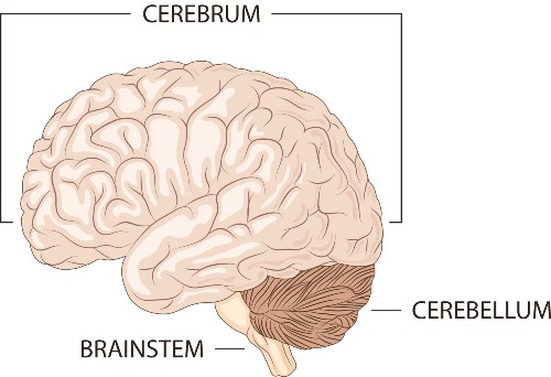
The brain is divided into three main parts:
The spinal cord is attached to the brain stem. The meninges is the layer of tissue covering the brain.

The cerebrum
The cerebrum is the front of the brain, and it is the largest part. It is divided into two hemispheres – the left hemisphere and the right hemisphere. The left hemisphere controls the right side of the body, and the right hemisphere controls the left side of the body. The two sides of the brain are also responsible for the following functions:
Left hemisphere
- Speaking
- Listening
- Reading
- Writing
Right hemisphere
- Following directions
- Solving puzzles
- Drawing
- Recognizing objects/people
The cerebrum is covered by the cerebral cortex. This is commonly called grey matter. It gives the brain a wrinkled appearance. Each fold in the grey matter is called a gyrus, and each valley between the folds is called a sulcus. These folds and valleys increase the number of neurons in the brain, meaning the brain can function at a higher level.
The four lobes of the brain
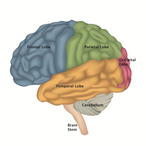
Within the cerebrum and the two hemispheres, there are four lobes of the brain that control different functions.

The parietal lobe is responsible for sending and receiving visual, auditory, and touch information. Anything we see, hear, or physically feel is processed in this part of the brain. The parietal lobe is also divided into a left and right side.
The left side is responsible for recognizing speech and words. Damage to the left parietal lobe may lead to problems in reading and math. It can also cause a loss of sensation on the right side of the body. This means that you would have changes in how you feel touch or pain, vision, or temperature changes on your right side.
The right parietal lobe is responsible for visual and spatial recognition. This includes shapes and body orientation when moving around. Damage to the right parietal lobe may lead to problems with spatial tasks, like making sense out of pictures, diagrams, and reading maps. Damage may also cause changes in sensation on the left side of the body.
The temporal lobe is responsible for distinguishing smells and sounds. It also completes the following jobs:
- Memory acquisition and storage
- Perception
- Categorization of objects
There are left and right sides in the temporal lobe.
The left temporal lobe is responsible for language comprehension through listening and reading. Damage to the left temporal lobe may cause problems in understanding and remembering language.
The right temporal lobe helps you comprehend non-verbal sounds (like a car horn) and music. Damage to the right temporal lobe may cause problems in understanding and remembering non-verbal information such as pictures, diagrams, body language cues, and other visual messages.
The occipital lobe processes visual information like patterns and colours. This helps you identify objects and signals. Damage to the left occipital lobe may cause problems seeing things on the right side of space. Damage to the right occipital lobe may cause problems seeing things on the left side of space.
The frontal lobe is the decision-making centre of your brain (also called the executive function centre). It receives information from the other lobes and figures out the best way to respond to interact with the environment. The frontal lobe controls the following things:
- Memory
- Concentration
- Judgement and inhibition
- Movement
- Smell
- Taste
- Self-awareness
- Personality traits
- Organization
- Word formation
Damage to either side of the frontal lobe may lead to problems with emotional control, social skills, judgment, planning, and organization.
Damage to the left frontal lobe may cause problems with speech and moving the right arm or leg.
Damage to the right frontal lobe may cause problems moving the left arm or leg.
Cortical and subcortical structures
Diving deeper into how the brain works, there are cortical and subcortical structures that have more specific functions.
The basal ganglia is a group of neurons located at the base of the cerebrum. It is responsible for gross motor function, posture, and balance. It also helps with automatic learning of movements used without conscious thought, like walking, biking, or picking things up.
Damage to the basal ganglia results in difficulty moving, or an inability to perform some of these basic automatic actions.
The hypothalamus is a small section at the base of the brain that controls the nervous system through hormone production and release. It is responsible for regulating things like:
- Hunger
- Thirst
- Blood pressure
- The sleep/wake cycle
- Sexual arousal
Damage to the hypothalamus can result in lack of motivation, changes in thirst/hunger routines, feelings of euphoria, hyperactivity, or altered sleep patterns.
The thalamus is the central relay station for incoming sensory information. Every sensation you experience travels through the thalamus. It plays an important part in pain information, memory, alertness and attention span.
Damage to the thalamus can result in many symptoms, including trouble processing information and spatial neglect.
The limbic system is responsible for all brain structures involved in emotion, motivation, and association with memory. This includes remembering emotional states and physical sensations. Damage to the limbic system can affect your ability to process and store information, as well as cause disruptions in emotion. Within the limbic system are several structures.
The parahippocampal gyrus is responsible for spatial memory formation. This means you remembering where objects are or where things happen in proximity to the body.
The cingulate gyrus controls autonomic functions like heart rate regulation. It also controls cognitive and attentional processing.
The nucleus accumbens mediates reward-based feelings. It also controls sensations associated with addiction.
This pea-sized part of the limbic system receives smell inputs and transmits them using olfactory tracts to other parts of the brain. It helps identify smells.
This is the largest white matter portion of the brain. It is an area of nerve cells located between the right and left hemispheres, and it allows them to communicate with each other. Cognitive, motor, and sensory information can be shared between the two hemispheres. Through these connections, we experience connected vision and connect objects with language.
There are two mammillary bodies, and they help pass information to other parts of the brain that are in charge of creating memories.
The hippocampus is responsible for creating long-term memory. Any damage to this area of the brain would result in difficulty or the inability to create memories.
The fornix connects the hippocampus and the mammillary bodies. The hippocampus uses the fornix to transmit information, which the mammillary bodies receive information.
The amygdala processes feelings like love, aggression and fear, and the responses of the body when you experience those feelings. It also helps us recognize those emotions in other people.
The stria terminalis connects the amygdala to the hypothalamus along the surface of the thalamus. It helps regulate responses to stress in its role as an output path from the amygdala.
The cerebellum
The cerebellum is located at the base of the back of the brain and integrates sensory perception and motor output. This includes:
- Balance
- Coordination
- Posture
- Speech
Connected to multiple parts of the brain, the cerebellum coordinates voluntary movements. This results in a smooth execution process when it comes to things like walking, speaking, and smaller movements like lifting and reaching. Damage to the cerebellum may cause problems with coordination, balance, or muscle tone in different parts of the body.
The brainstem
The brainstem connects the brain to the spinal cord, and relays information back and forth from the brain to the peripheral nervous systems. The brainstem controls basic life functions, including:
- Breathing
- Blood pressure
- Heart rate
- Sweating
- Digestion
- Arousal
- Alertness
There are three main parts of the brainstem.
The midbrain is responsible for auditory and visual reflexes. It controls eye movements, processes sight and sounds and how your eyes and ears react to the environment.
Pons is the largest part of the brain stem. It acts as a bridge between the cerebrum and cerebellum. The pons controls various parts of the body, including facial nerves and movements. One of its most important roles is as a respiratory information centre. The pons controls breathing, including information on carbon dioxide and oxygen levels.
The medulla oblongata also helps control respiration through controlling the heart and lungs. It is responsible for vasomotor actions, cardiac functions and reflex actions like sneezing or coughing. Any damage to the brain stem can cause physical and sensory problems. This includes:
- Heartbeat and breathing can be disrupted or stopped
- Swelling leading to hemorrhaging and stroke
- Speech impairment
- Difficulty swallowing
- Personality changes
Memory loss
In the most severe cases, damage to the brainstem can lead to coma or paralysis.
This is a basic overview of parts of the brain and how they work. If you have more specific questions, ask to speak to a neuropsychologist.
Definitions and sources
Auditory (au-di-tor-y): your sense of hearing.
Cortical (cor-ti-cal): relating to the outer layer of the cerebrum.
Grey matter (grey mat-ter): the darker tissue of the brain and spinal cord.
Gyrus (gy-rus): a ridge or fold between two clefts on the surface of the brain.
Hemisphere (hem-i-sphere): half of a sphere or three-dimensional circle.
Lobes: any part of an organ that seems to be separate in some way from the rest.
Motor function (mo-tor func-tion): any act or movement which is completed using motor neurons. Mo-tor neu-rons are a nerve cell that helps form pathways that passes from the brain or spinal cord to a muscle or gland.
Nerve tissue (nerve tis-sue): made up of neurons and supporting cells. One of the main four
types of tissue in your body.
Neurons (neu-rons): a specialized cell transmitting nerve impulses. They transmit information throughout the body.
Peripheral nervous system (pe-riph-er-al nerv-ous sys-tem): the nervous system outside the brain and spinal cord.
Sensory (sen-so-ry): what you experience through the physical senses of touch, sight, sound, taste, or smell.
Spatial (spa-tial): relating to or occupying space.
Subcortical (sub-cor-ti-cal): the area of the brain below the cortex.
Sulcus (sul-cus): a groove or furrow on the surface of the brain.
All definitions are taken or adapted from dictionary.com
Information for this page provided in part by Ontario Brain Injury Association and My Health Alberta.
Disclaimer: There is no shortage of web-based online medical diagnostic tools, self-help or support groups, or sites that make unsubstantiated claims around diagnosis, treatment and recovery. Please note these sources may not be evidence-based, regulated or moderated properly and it is encouraged individuals seek advice and recommendations regarding diagnosis, treatment and symptom management from a regulated healthcare professional such as a physician or nurse practitioner. Individuals should be cautioned about sites that make any of the following statements or claims that:
- The product or service promises a quick fix
- Sound too good to be true
- Are dramatic or sweeping and are not supported by reputable medical and scientific organizations.
- Use of terminology such as “research is currently underway” or “preliminary research results” which indicate there is no current research.
- The results or recommendations of product or treatment are based on a single or small number of case studies and has not been peer-reviewed by external experts
- Use of testimonials from celebrities or previous clients/patients that are anecdotal and not evidence-based
Always proceed with caution and with the advice of your medical team.